cortical-depth-dependent vascular responses in cerebral amyloid angiopathy

Problem

There has been research to prove that cerebral amyloid angiography (CAA) can lead to measurable differences in the fMRI response amplitude.

Objective

For this study, the goal was to examine the hemodynamic responses across different cortical depths driven by visual stimulation and CO2 inhalation.

Skills Employed

  • MATLAB
  • Linux environment
  • Statistical analysis and basic signal processing
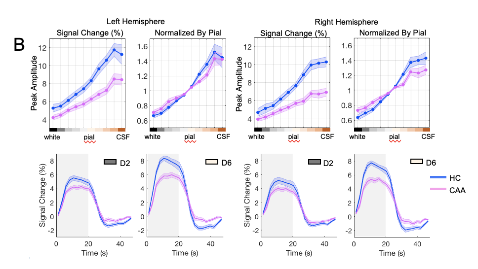
Image of CAAfig2B
Figure 1. Graphs showing the percent signal change of the brain at different depths of the left and right hemisphere.

Approach

Essentially:

  • Estimated vascular responses driven by both visual and physiological stimulus
  • Characterized the progression patterns of vascular responses across cortical depths
  • Compared the derived vascular response patterns between CAA vs. healthy subjects
  • Co-authored conference abstract of the study findings submitted to the Organization of Human Brain Mapping 2021 conference
  • Attended the Organization of Human Brain Mapping 2021 Virtual conference


Image of CAAfig2B
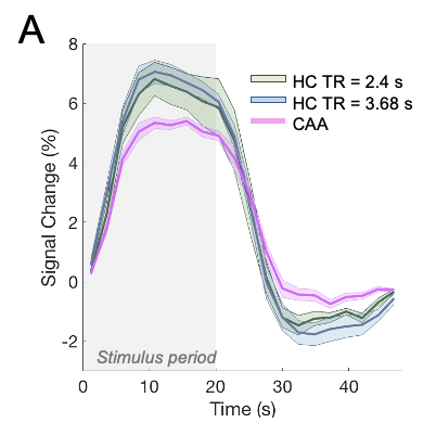
Figure 2. Graph showing the percent signal change of the brain of heathy subjects (at two different MRI repetition times (TR)) and of subjects with cerebral amyloid angiopathy (CAA)

Results

The developed MATLAB code successfully examined the hemodynamic responses across different cortical depths driven by visual stimulation and CO2 inhalation and resulted in a conference abstract!

to the top
research
the whole shabang

Copyright © Jen Yeo 2024